
Every intricate twist, powerful leap, and delicate touch is a testament to the remarkable engineering within our bodies, none more so than our tendons. These robust, yet often overlooked, fibrous cords act as the essential bridge between muscle and bone, seamlessly converting muscular contraction into graceful, purposeful motion. Despite their intrinsic strength and critical role in daily life and athletic endeavors, tendon injuries are incredibly common, frequently causing persistent pain, limiting activity, and impacting overall quality of life.
Are you struggling with that nagging ache, stiffness, or sharp pain that just won’t go away? You’re likely experiencing a tendon injury. Understanding these vital biological structures is the bedrock of effective recovery and sustained well-being. This comprehensive guide is your definitive resource, designed to equip you with world-class insights. We’ll meticulously explore the anatomy of tendons, dissect the spectrum of common tendon injuries, unveil cutting-edge prevention strategies, and outline the most effective, evidence-based methods to treat tendon injuries and heal tendon injuries swiftly and without lingering discomfort. Prepare to reclaim a pain-free, active life by mastering the art of tendon health.
Understanding Tendons: The Unsung Architects of Movement
To truly conquer tendon injuries, we must first gain a profound appreciation for the intricate design and paramount function of tendons. These robust structures are absolutely fundamental to our capacity for movement, stability, and athletic prowess.
What Are Tendons? Definition & Function Beyond the Basics
Tendons are complex biological marvels, appearing as tough, exquisitely organized bands of fibrous connective tissue. Primarily composed of Type I collagen fibers, they are arranged in a hierarchical fashion: tiny fibrils bundle into fascicles, which then form the complete tendon. Imagine them as high-tensile, multi-stranded ropes, engineered for immense tensile strength, allowing them to withstand significant pulling forces generated by muscle contractions without yielding.
Key components that contribute to the remarkable properties of tendons include:
- Collagen Fibers (Type I): The architectural backbone, providing unparalleled tensile strength and structural integrity. These fibers are highly ordered and aligned along the direction of mechanical stress.
- Elastin: Present in smaller quantities, offering a crucial degree of elasticity, enabling some stretch and recoil and helping dissipate forces.
- Ground Substance: A gel-like matrix of glycoproteins and proteoglycans. This substance organizes collagen fibers, maintains tissue hydration, and plays a role in mechanotransduction (how cells sense and respond to mechanical stimuli).
- Tenocytes and Tenoblasts: These specialized cells reside within the tendon matrix. Tenoblasts are immature, metabolically active cells responsible for matrix synthesis and repair. They mature into tenocytes, which are elongated cells embedded within the collagen bundles, maintaining the tendon’s structure and responding to mechanical loads.
- Mitochondrial Network: Crucial for energy production, especially in highly active tendons, supporting continuous repair and remodeling processes.
When a muscle contracts, it generates force. This force is efficiently and directly transmitted through the tendon to its attached bone, initiating movement. Tendons act as biological levers, converting muscle power into skeletal movement. Their relative stiffness compared to muscle tissue makes them exceptional transmitters of force, while their viscoelastic properties allow them to absorb, store, and release elastic energy, making activities like jumping, running, and throwing more efficient. This energy storage capacity reduces the metabolic cost of movement and protects muscles from excessive strain, playing a vital role in preventing certain tendon injuries.
Tendons vs. Ligaments: A Critical Distinction for Injury Management
While often confused due to their similar appearance and connective tissue classification, tendons and ligaments serve fundamentally distinct purposes. This is a crucial distinction for accurate diagnosis and effective strategies to treat tendon injuries.
- Tendons: Connect muscle to bone. Their primary and most vital role is to facilitate movement by transmitting muscular force and to absorb shock during dynamic activities.
- Ligaments: Connect bone to bone. Their main function is to stabilize joints, preventing excessive or unwanted movement, thereby protecting joint structures.
Understanding this fundamental difference is not just academic; it dictates the approach to diagnosis and the specific strategies employed for treating tendon injuries versus ligamentous sprains. Injuries to ligaments (sprains) typically involve joint instability, whereas injuries to tendons (tendinopathies) primarily affect force transmission and movement execution.
Key Tendons & Their Vulnerabilities: Identifying High-Risk Areas
From the simplest act of maintaining posture to executing a sophisticated athletic maneuver, tendons are in constant engagement, subjected to varying degrees of stress. Their specific location and function often dictate their unique vulnerabilities, making certain areas common sites for tendon injuries.
- Achilles Tendon: The largest and strongest tendon in the human body, connecting the calf muscles to the heel bone. It’s indispensable for walking, running, jumping, and virtually all propulsion activities. An Achilles tendon injury, ranging from tendinopathy to a complete rupture, can be severely debilitating, often requiring extensive rehabilitation or surgical intervention. Its vulnerability stems from the immense forces it endures, particularly during explosive movements.
- Rotator Cuff Tendons: A crucial group of four tendons (Supraspinatus, Infraspinatus, Teres Minor, Subscapularis) in the shoulder that surround the shoulder joint. They enable a wide range of arm movements (lifting, rotation) and provide critical stability. This area is a very common site for overuse tendon injuries, often leading to impingement, tendinopathy, or tears, particularly in overhead athletes or those performing repetitive arm movements.
- Patellar Tendon: Connects the quadriceps muscle (via the patella) to the shin bone. Absolutely essential for knee extension and activities involving jumping, kicking, and running. Often associated with “jumper’s knee” (patellar tendinopathy), this tendon injury arises from repetitive loading and explosive forces, common in sports like basketball and volleyball.
- Common Extensor & Flexor Tendons (Elbow): The common extensor tendon, attaching to the lateral epicondyle, is the site of “tennis elbow” (lateral epicondylitis/tendinopathy), affecting forearm extensors. The common flexor tendon, attaching to the medial epicondyle, is the site of “golfer’s elbow” (medial epicondylitis/tendinopathy), affecting forearm flexors. Both are classic examples of overuse tendon injuries from repetitive gripping, wrist extension/flexion, and arm movements.
- Wrist and Hand Tendons: A multitude of smaller, intricate tendons facilitate the fine, precise movements of our fingers and hands. Their complex routing and constant activity make them highly susceptible to repetitive strain tendon injuries, such as De Quervain’s tenosynovitis or trigger finger, often seen in occupations requiring fine motor skills or prolonged gripping.
- Gluteal Tendons (Gluteus Medius and Minimus): Located around the hip, these tendons are critical for hip abduction and pelvic stability. Tendinopathy in these tendons is a common cause of lateral hip pain, particularly in runners and older adults.
Maintaining optimal tendon health across all these diverse anatomical areas is not merely beneficial but vital for an active, functional, and pain-free life.
While we often focus on major tendon groups, even smaller ones can be susceptible to injury, highlighting the importance of proper ergonomics and addressing conditions like De Quervain’s Syndrome with supportive medical devices to prevent further complications.
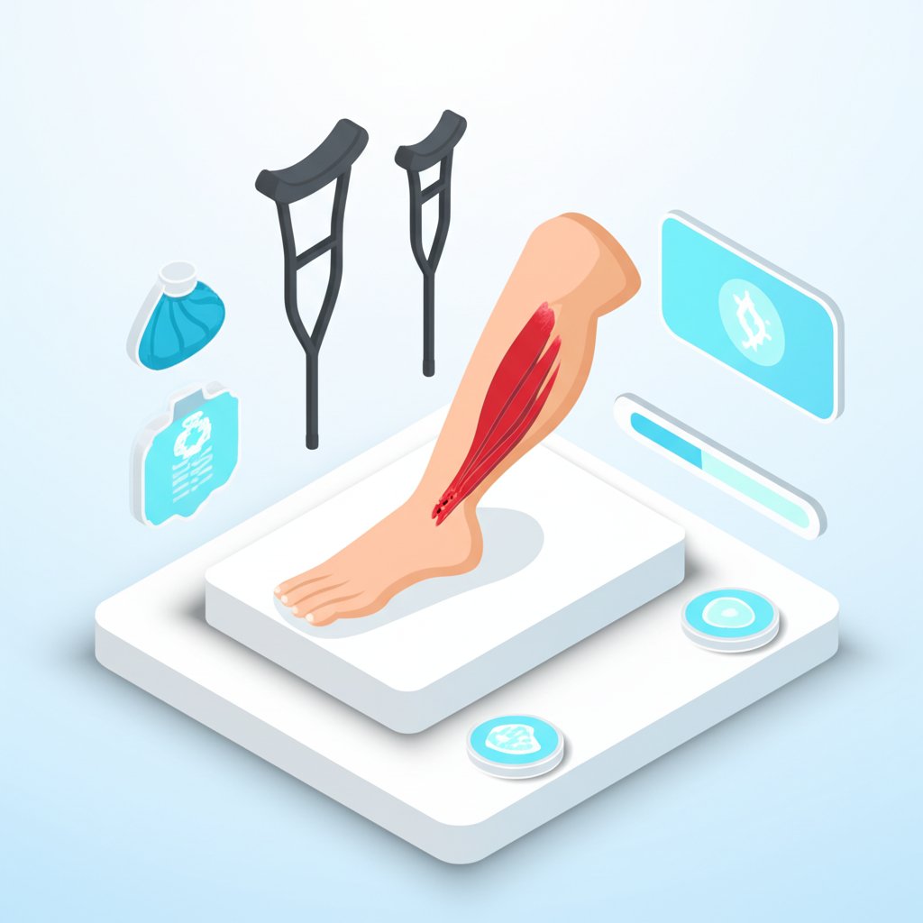
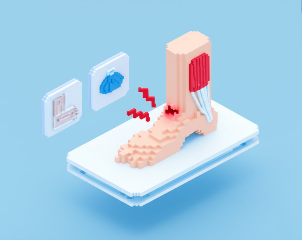
Decoding Tendon Injuries: Types, Causes, and Critical Symptoms
Tendon injuries, broadly categorized as tendinopathies, represent a widespread musculoskeletal problem, impacting individuals across all age groups and activity levels. These injuries most frequently stem from a combination of repetitive strain, sudden traumatic incidents, or insidious age-related degeneration. Accurately understanding the specific type of tendon injury is the cornerstone of effective management and successful long-term recovery.
Differentiating Tendinitis (Acute) from Tendinosis (Chronic Degeneration)
Historically, the vast majority of tendon injuries were generically labeled as “tendinitis,” implying an inflammatory process. However, modern research has refined our understanding.
- Tendinitis: Technically refers to an acute inflammatory response within the tendon, typically occurring in response to a sudden, unaccustomed overload, a sharp increase in activity, or an acute traumatic event causing micro-tears.
- Symptoms: Sharp, localized pain (often immediate and worsens acutely with movement), swelling, tenderness to the touch, warmth, and sometimes redness.
- Reality Check: While acute inflammation can occur, particularly in the initial 24-72 hours post-injury, contemporary research and histological studies now reveal that chronic tendon injuries (those lasting more than 6-12 weeks) rarely exhibit significant numbers of inflammatory cells. This crucial discovery has revolutionized our approach to chronic tendinopathy.
- Tendinosis: Now recognized as a far more common and often chronic form of tendon injury, characterized by degenerative changes within the tendon’s internal structure rather than primary inflammation. It results from a mismatch between tendon loading and its capacity for repair – repeated micro-traumas accumulate, and the tendon’s intrinsic repair mechanisms cannot keep pace with the ongoing damage. This leads to structural breakdown.
- Key Pathological Features: Disorganized collagen fibers, increased ground substance, neovascularization (new, often immature, blood vessels), neuronal ingrowth (new nerve endings correlating with persistent pain), and an absence of significant inflammatory cells.
- Symptoms: Typically more chronic, dull, and aching pain. It often worsens predictably with specific activities, improves somewhat with rest, but consistently returns upon re-engagement in the aggravating activity.
- Clinical Importance: It’s paramount to differentiate tendinitis from tendinosis, as their strategies to treat tendon injuries diverge significantly – anti-inflammatory approaches are less effective for tendinosis, which requires load-based rehabilitation.
Tenosynovitis & Paratenonitis: Inflammation of the Tendon Sheath
Certain tendons, particularly in areas prone to high friction or repetitive gliding (e.g., wrist, hand, ankle, foot), are enveloped by a protective synovial sheath. This sheath contains synovial fluid, which lubricates the tendon, allowing it to glide smoothly.
- Tenosynovitis: The inflammation of this synovial sheath, often caused by repetitive friction or compression.
- Symptoms: Localized pain and tenderness along the tendon’s actual path (often worse with movement), swelling, a creaking or grating sensation (crepitus) when moving the affected tendon, and limited range of motion.
- Example: De Quervain’s tenosynovitis, affecting the thumb side of the wrist (abductor pollicis longus and extensor pollicis brevis), common in new mothers or those performing repetitive gripping.
- Paratenonitis: Inflammation of the paratenon, the delicate tissue layer surrounding tendons that do not have a synovial sheath (e.g., Achilles tendon). It acts similarly to a sheath, reducing friction.
- Symptoms: Similar to tenosynovitis, including pain, tenderness, and sometimes crepitus, but without true sheath inflammation.
Partial vs. Complete Tendon Ruptures: Recognizing the Severity
A tendon rupture represents the most severe end of the tendon injury spectrum, involving a partial or complete tearing of the tendon fibers. These catastrophic injuries typically occur due to a sudden, forceful contraction of the muscle against strong resistance or an acute traumatic event, often when the muscle is already under tension.
- Partial Rupture: Involves some fibers being torn, but the tendon largely remains intact.
- Symptoms: Sudden, sharp, often severe pain; swelling and bruising; weakness and difficulty performing actions that utilize the injured tendon, though some function may remain; localized tenderness; a feeling of a “pop” or “snap” may or may not be heard.
- Complete Rupture: The tendon is fully severed, creating a complete loss of continuity between the muscle and the bone, rendering it unable to transmit force.
- Symptoms: An unmistakable, loud “pop” or “snap” sound at the moment of injury, often described as feeling like being hit by a golf ball; intense, excruciating pain (though sometimes initial pain quickly subsides, followed by a dull ache); immediate and profound loss of function (e.g., inability to push off the foot with an Achilles rupture); a visible deformity or palpable gap where the tendon should be; significant swelling and bruising.
- Common Sites: Achilles tendon, rotator cuff tendons, quadriceps tendon, patellar tendon, and biceps tendon. These severe tendon injuries almost always necessitate urgent medical assessment and frequently require advanced medical interventions, including surgical repair, to restore function.
Risk Factors & Why Some Are More Susceptible
Several intrinsic and extrinsic factors collectively contribute to an individual’s vulnerability to tendon injuries. Understanding these allows for targeted prevention.
- Age: A significant intrinsic factor. As we age, tendons naturally undergo degenerative changes: they become thinner, blood flow decreases, collagen synthesis slows, and tenocyte activity diminishes. Collagen fibers accumulate damage, becoming more cross-linked and stiffer. These changes reduce elasticity and resilience, making the tendon significantly more susceptible to tendinosis and acute ruptures, particularly after age 30-40.
- Overuse and Repetitive Strain: The single most common extrinsic cause. Activities involving highly repetitive movements or sustained loading without adequate recovery time can cumulatively exceed the tendon’s adaptive capacity, leading to micro-trauma and the development of tendinopathy.
- Poor Biomechanics or Technique: Incorrect form or inefficient movement patterns during exercise, sports, or daily activities can place undue, asymmetrical, or excessive stress on specific tendons. This leads to localized overload and injury, even if overall training volume isn’t high.
- Sudden Increase in Activity (“Too Much, Too Soon”): Rapidly increasing the intensity, duration, or frequency of exercise without allowing tendons sufficient time to adapt and strengthen is a prime recipe for tendon injuries. Tendons adapt slower than muscles.
- Inadequate Warm-up or Cool-down: Skipping these steps leaves tendons unprepared for stress or impairs their ability to flush metabolic waste and initiate repair processes.
- Muscle Imbalances: Weakness, tightness, or altered activation patterns in surrounding muscles can disrupt joint mechanics, leading to abnormal loading and compensation that place excessive strain on specific tendons.
- Certain Medical Conditions:
- Diabetes: Can lead to advanced glycation end products (AGEs) in collagen, making tendons stiffer, less resilient, and impairing blood flow and healing.
- Rheumatoid Arthritis and other Autoimmune Diseases: Can cause inflammation that directly affects tendons and sheaths.
- Thyroid Disorders: Hypothyroidism is associated with increased risk of tendinopathy and rupture.
- Medications:
- Fluoroquinolone Antibiotics (e.g., Ciprofloxacin, Levofloxacin): Notorious for increasing the risk of tendinopathy and, more severely, tendon rupture (especially Achilles), sometimes even weeks or months after treatment.
- Corticosteroids (oral or injected): While short-term anti-inflammatory, repeated local injections can weaken tendon tissue and increase rupture risk.
- Poor Nutrition: Chronic lack of essential nutrients can impair the tendon’s ability to synthesize new collagen and repair itself efficiently.
- Obesity: Increased body weight places greater mechanical load on weight-bearing tendons, increasing the risk of injury.
- Smoking: Nicotine negatively impacts blood flow and tissue healing, reducing the tendon’s capacity for repair.
Accurate Diagnosis: The First Step to Effective Treatment
When a tendon injury is suspected, a precise diagnosis is crucial for guiding effective treatment and optimizing the healing process.
- Comprehensive Medical History: Your doctor will inquire about the onset of pain, specific activities that worsen or improve it, previous injuries, your activity level, occupation, and any relevant medical conditions or medications (like fluoroquinolone antibiotics).
- Detailed Physical Examination: This is paramount. The clinician will palpate the affected tendon for tenderness, swelling, and any palpable gaps (indicating rupture). They will assess your range of motion, strength, and perform specific orthopedic tests to identify the injured tendon and differentiate it from other issues like ligament sprains or muscle strains. For example, specific resistance tests can isolate individual rotator cuff tendons.
- Diagnostic Imaging:
- X-ray: Primarily used to rule out bone fractures or avulsion injuries (where a piece of bone pulls away with the tendon). It doesn’t show soft tissues well.
- Ultrasound: A dynamic, non-invasive, and cost-effective imaging tool. It can effectively visualize tendon integrity, detect thickening, tears (partial or complete), neovascularization (common in tendinosis), and inflammation of the tendon sheath (tenosynovitis). It allows for dynamic assessment of the tendon during movement.
- Magnetic Resonance Imaging (MRI): Considered the gold standard for soft tissue evaluation. MRI provides detailed images of the tendon’s internal structure, surrounding soft tissues, and bone. It is excellent for confirming the extent of partial tears, precisely identifying complete ruptures, and evaluating chronic tendinopathy changes (like collagen disorganization and mucoid degeneration). It also helps rule out other causes of pain.
- Computed Tomography (CT) Scan: Less commonly used for primary tendon injuries, but can be helpful for assessing bony involvement or complex joint anatomy, especially if surgery is being considered in areas with complex bone structures.
A thorough diagnostic process ensures that the specific nature and severity of your tendon injury are understood, allowing for a tailored and effective treatment plan.
Comprehensive Strategies to Treat Tendon Injuries Effectively
Successfully navigating a tendon injury requires a multi-faceted approach, moving beyond simple rest to embrace active, evidence-based interventions. The goal is not merely to alleviate pain but to restore the tendon’s structural integrity, strength, and function, allowing you to heal tendon injuries completely and return to desired activities.
Immediate First Aid: The R.I.C.E. Principle & Beyond
For acute tendon injuries (e.g., sudden onset pain, swelling), immediate first aid can significantly impact initial pain and swelling.
- R – Rest: Crucial in the early stages. Avoid activities that aggravate the injured tendon. For severe injuries, immobilization (e.g., brace, walking boot) may be necessary. However, prolonged, complete rest should be avoided for tendinopathy, as it can be detrimental to long-term healing.
- I – Ice: Apply a cold pack to the affected area for 15-20 minutes, several times a day, especially in the first 24-48 hours. This helps reduce pain, swelling, and acute inflammation. Always place a thin cloth between ice and skin.
- C – Compression: Using an elastic bandage can help minimize swelling, but ensure it’s not too tight to impede circulation.
- E – Elevation: Elevating the injured limb above heart level (if feasible) also aids in reducing swelling.
- Beyond RICE: Consider gentle, pain-free movement within the first few days if it’s a mild injury. For chronic tendinopathy, ice may still help with pain, but the focus shifts rapidly to controlled loading.
Conservative Management: Physical Therapy, Orthotics & Manual Therapy
The vast majority of tendon injuries, especially tendinopathies, respond exceptionally well to conservative management, with physical therapy being the cornerstone.
- Physical Therapy (PT): A tailored PT program is essential to
treat tendon injuriesby improving strength, flexibility, and biomechanics.- Eccentric Loading: Often considered the gold standard for tendinopathy. These exercises involve lengthening the muscle under tension (e.g., the lowering phase of a calf raise for Achilles tendinopathy). Eccentric loading specifically stimulates collagen remodeling, increases the tendon’s load-bearing capacity, and reduces pain. Your therapist will guide you on appropriate intensity and volume.
- Isometric Exercises: Holding a muscle contraction at a fixed length (e.g., wall sit for patellar tendinopathy). These can be excellent for immediate pain relief and building foundational strength without causing joint movement, especially in acute phases.
- Progressive Resistance Training: Gradually increasing the load and complexity of exercises to rebuild strength in the muscle-tendon unit.
- Proprioception and Balance Training: Crucial for restoring coordinated movement and reducing recurrence risk, especially for lower limb tendon injuries.
- Stretching and Flexibility: Addressing muscle tightness that may be contributing to abnormal tendon loading.
- Orthotics and Bracing: Custom or off-the-shelf orthotics (e.g., shoe inserts) can correct foot biomechanics that place undue stress on Achilles or patellar tendons. Braces or splints can provide support and limit motion during the initial healing phases or during activity.
- Manual Therapy: Techniques like massage, soft tissue mobilization, and joint mobilizations performed by a physical therapist can help reduce pain, improve tissue extensibility, and address compensatory muscle tightness.
Pharmaceutical & Injection Therapies: What Works & What to Avoid
Medications can assist in managing pain and inflammation, but some carry risks, especially for tendons.
- NSAIDs (Non-Steroidal Anti-Inflammatory Drugs): Over-the-counter NSAIDs like ibuprofen or naproxen can help reduce pain and swelling, particularly in acute tendinitis. For chronic tendinosis, their anti-inflammatory effects are less relevant, but they can still provide short-term pain relief. Use judiciously and for short durations.
- Corticosteroid Injections: While offering rapid pain relief in some cases, corticosteroids can actually weaken tendon tissue and increase the risk of rupture, particularly with repeated injections. They are generally not recommended for chronic tendinosis and should be used with extreme caution and sparingly, if at all, for acute tendinitis.
- Topical Pain Relievers: Gels or creams containing NSAIDs can provide localized pain relief with fewer systemic side effects.
Advanced Non-Surgical Interventions: PRP, Shockwave & Dry Needling
For chronic tendon injuries that haven’t responded to conventional conservative treatment, several advanced non-surgical options are gaining traction.
- Platelet-Rich Plasma (PRP) Injections: PRP involves drawing a small amount of your blood, processing it to concentrate platelets (which contain growth factors), and then injecting this concentrated plasma into the injured tendon. The growth factors are believed to stimulate cell proliferation and tissue regeneration, promoting healing.
- Extracorporeal Shockwave Therapy (ESWT): ESWT delivers high-energy acoustic waves to the injured tendon. This is thought to stimulate healing by promoting neovascularization, breaking down calcifications (common in some chronic tendinopathies), and stimulating a healing response in chronic, degenerated tendons.
- Dry Needling: Involves inserting thin, sterile needles into trigger points or dysfunctional areas within the muscle or around the tendon. While not directly treating the tendon itself, it can alleviate muscle tightness and pain that contribute to tendon overload.
- Sclerosing Injections: Injections of hyperosmolar dextrose or polidocanol aim to sclerose (harden) abnormal neovessels and nerve ingrowth found in chronic tendinosis, which are thought to be sources of chronic pain.
These advanced treatments are typically considered after a trial of physical therapy and should be discussed thoroughly with a specialist, weighing potential benefits against risks and costs.
When is Surgery Necessary for Tendon Injuries?
Surgery is generally reserved for specific types of tendon injuries or when conservative measures have failed after an extended period (e.g., 6-12 months) of dedicated effort.
- Complete Tendon Ruptures: Most complete ruptures, especially in major tendons like the Achilles, quadriceps, or rotator cuff, require surgical repair to restore function. The goal is to reattach the torn ends of the tendon or reattach the tendon to the bone.
- Large Partial Tendon Tears: Depending on the size, location, and functional impact, large partial tears, particularly in the rotator cuff, may benefit from surgical repair to prevent progression to a complete rupture and restore strength.
- Chronic Tendinopathy Resistant to Conservative Treatment: In rare cases of severe, chronic tendinosis with significant structural degeneration and persistent pain that has failed all non-surgical interventions, surgical debridement (removing damaged tissue) or repair may be considered.
- Tenosynovitis with Mechanical Impingement: If conservative treatments fail to relieve symptoms in conditions like trigger finger or De Quervain’s tenosynovitis, a surgical release of the tendon sheath may be performed to decompress the tendon.
Surgical intervention carries its own risks and requires a dedicated rehabilitation period. The decision for surgery should always be made in consultation with an orthopedic surgeon, considering the individual’s activity level, age, overall health, and specific injury characteristics.
Accelerating Healing & Recovery: How to Heal Tendon Injuries Faster
The journey to heal tendon injuries is a process that demands patience and a strategic approach.