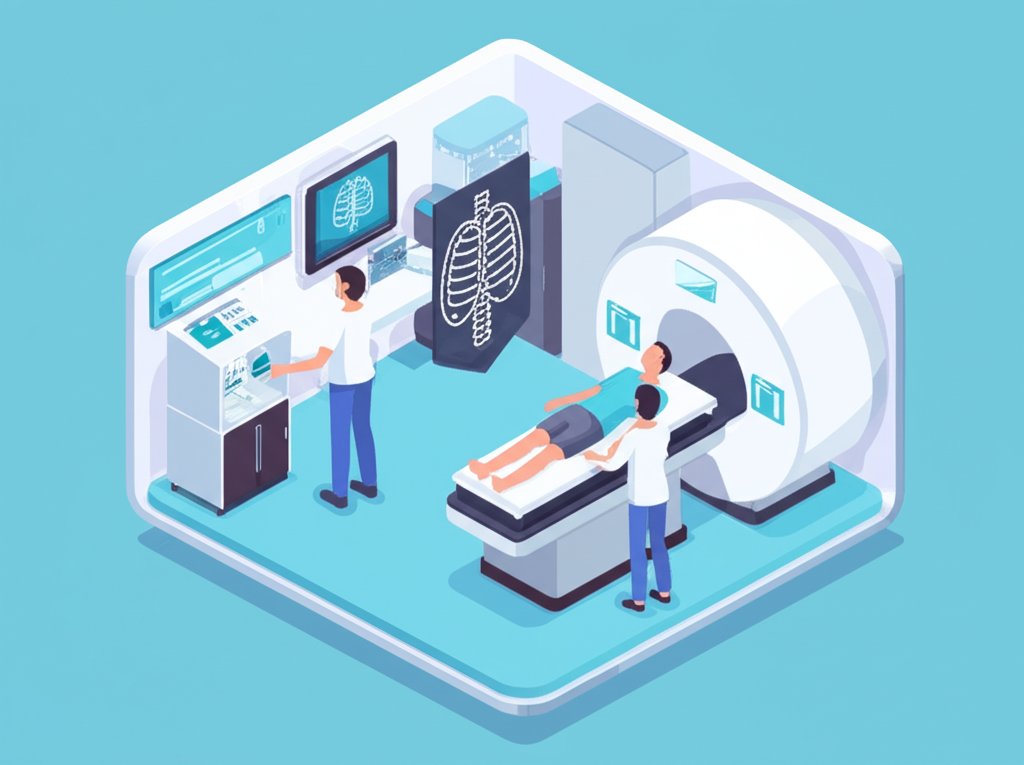
In an era defined by rapid medical advancements, the landscape of healthcare is constantly evolving, driven by an unyielding quest for safer, more effective, and patient-friendly solutions. At the forefront of this revolution stands non-invasive medical imaging – a sophisticated realm of medical imaging technology that has profoundly reshaped our approach to diagnosis, treatment, and overall patient care. Gone are the days when peering inside the human body inherently meant invasive procedures; today, advanced imaging techniques offer unparalleled views without a single incision, minimizing discomfort and risk while maximizing diagnostic precision.
This article delves into the intricate world of non-invasive medical imaging, exploring its fundamental principles, cutting-edge technologies, and its transformative impact on patient care. We will uncover how these remarkable innovations are not just improving current practices but are actively charting the course for the future of medicine, promising a healthier, more informed tomorrow.
Understanding Non-Invasive Medical Imaging: A Paradigm Shift
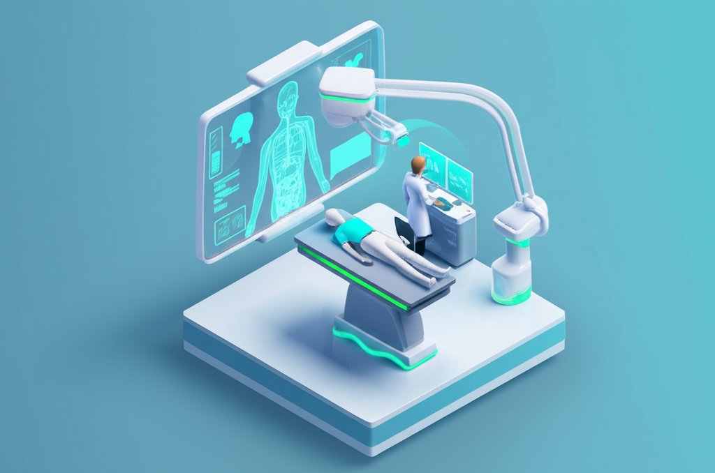
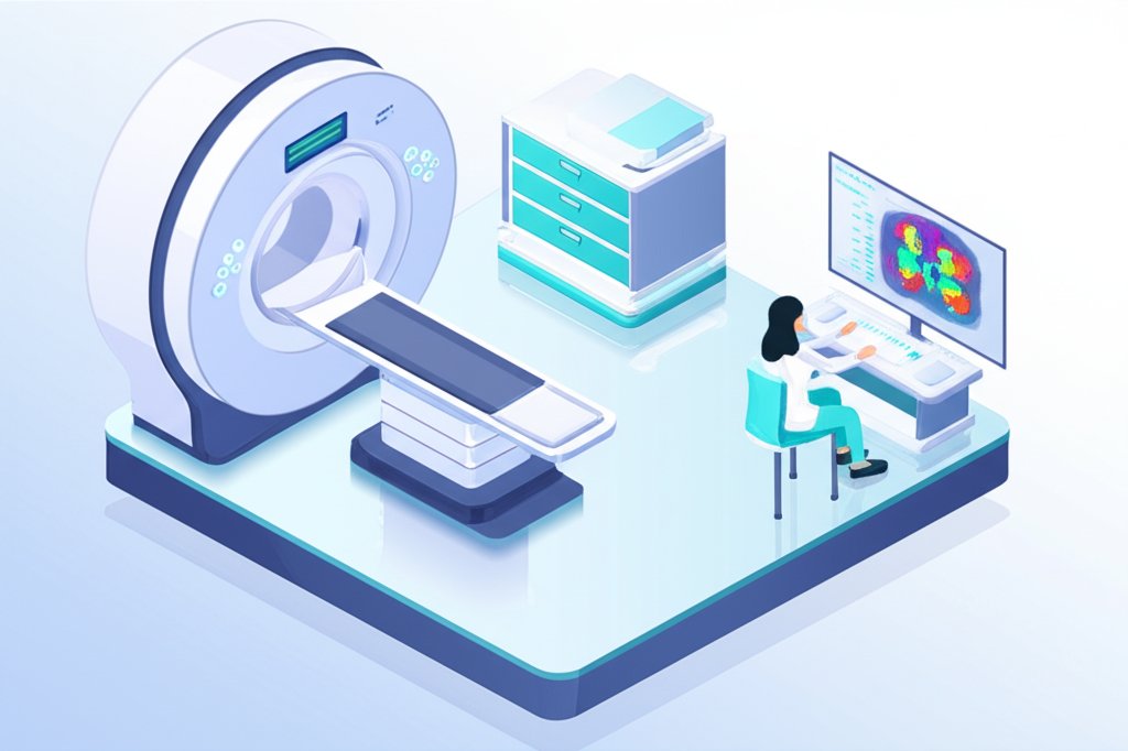
The concept of non-invasive medical imaging represents a monumental leap in diagnostic capabilities. By utilizing various forms of energy – from sound waves and magnetic fields to X-rays and radiotracers – these technologies create detailed images of internal body structures, functions, and even molecular processes, all without penetrating the skin or requiring surgery. This fundamental shift from intrusive exploration to external visualization has become a cornerstone of modern healthcare.
Defining the Non-Invasive Advantage: Beyond Procedures
The primary advantage of non-invasive procedures is inherent in their name: they avoid physical intrusion. This translates directly into numerous benefits for patients and healthcare systems alike:
- Reduced Risk: Eliminating the need for surgical incisions significantly lowers the risk of infection, bleeding, and complications associated with invasive interventions.
- Minimized Discomfort and Pain: Patients experience little to no pain during most non-invasive scans, enhancing their overall experience and reducing anxiety.
- Faster Recovery Times: Without surgical recovery, patients can typically resume normal activities immediately after an imaging session.
- Repeatability and Monitoring: The low-risk nature allows for repeated examinations, essential for monitoring disease progression, treatment response, and chronic conditions over time.
- Accessibility and Convenience: Many non-invasive medical imaging techniques are performed in outpatient settings, making them more accessible and convenient.
The Fundamental Principles: How We See Inside
At its core, medical imaging technology harnesses different physical principles to gather data about the body. Each modality offers a unique window:
- Computed Tomography (CT): Uses X-rays and computer processing to create cross-sectional images (slices) of bones, blood vessels, and soft tissues. It’s excellent for quickly detecting internal injuries and bone fractures.
- Magnetic Resonance Imaging (MRI): Employs powerful magnetic fields and radio waves to generate highly detailed images of soft tissues, organs, bone marrow, and virtually all internal structures. It’s invaluable for brain, spinal cord, and joint imaging.
- Ultrasound (US): Relies on high-frequency sound waves that bounce off internal structures to create real-time images. It’s commonly used for fetal imaging, cardiac evaluations, and abdominal assessments, notably with advancements like superb microvascular imaging.
- Positron Emission Tomography (PET): Uses small amounts of radioactive tracers to visualize metabolic activity and blood flow, often fused with CT scans (PET/CT) for precise anatomical localization. It’s crucial for oncology and neurology.
- Photoacoustic Imaging (PAI): A hybrid technique that uses light to generate ultrasound waves, offering high-resolution imaging with detailed functional and molecular information, particularly for vascular structures and tumor detection.
These diverse methods provide a comprehensive toolkit for medical professionals, allowing them to select the most appropriate imaging strategy based on the patient’s condition and diagnostic needs.
To get a clearer picture of your health status using some of these innovative tools, consider exploring the range of options available, including a comprehensive medical systems test menu to guide your diagnostic journey.
Core Technologies Driving Modern Medical Imaging
The rapid evolution of medical imaging technology is a testament to ongoing innovation, constantly pushing the boundaries of what’s possible in diagnostics. Beyond the foundational techniques, a new wave of advancements is enhancing clarity, speed, and diagnostic power.
Structural Insights: CT, MRI, and Ultrasound Enhancements
While CT, MRI, and ultrasound have been mainstays for decades, continuous innovation makes them even more powerful:
- Advanced CT Scans:
- Photon Counting CT (PCCT): This revolutionary technology directly detects individual X-ray photons, delivering higher spatial resolution, reduced noise, and the ability to distinguish different materials within the body more precisely, potentially reducing radiation dose.
- Deep-Learning Reconstruction: AI algorithms are used to reconstruct images from raw data, significantly improving image quality, reducing noise, and enabling faster scan times while maintaining diagnostic accuracy.
- Virtual Non-Contrast Imaging: Using advanced reconstruction, contrast-enhanced CT scans can generate “virtual” non-contrast images, reducing the need for an additional non-contrast scan and minimizing radiation exposure.
- Next-Generation MRI Techniques:
- Low-Field MRI: Moving away from ultra-high magnetic fields, low-field MRI systems offer more accessible, potentially portable, and less expensive options, while still providing valuable diagnostic information, especially in emergency settings or remote areas.
- MR Fingerprinting (MRF): This quantitative imaging technique rapidly acquires multiple tissue properties simultaneously, providing a more detailed and objective characterization of tissues than traditional MRI, aiding in early disease detection and treatment evaluation.
- Cutting-Edge Ultrasound:
- Superb Microvascular Imaging (SMI): A sophisticated Doppler technique that visualizes extremely slow blood flow in microvessels, often missed by conventional ultrasound, proving particularly valuable in tumor assessment and inflammatory conditions.
- Ultrafast Doppler Imaging: Enables real-time measurement of blood flow velocities at unprecedented speeds, offering new insights into cardiovascular dynamics.
- Ultrasound Localization Microscopy: A super-resolution imaging method that maps microvasculature with resolutions far beyond conventional ultrasound, akin to optical microscopy but deep within tissues.
- Magnetomotive Ultrasound (MMUS): Combines magnetic nanoparticles with ultrasound to detect and image specific molecular targets, enhancing diagnostic specificity.
Functional & Molecular Windows: PET and Specialized Techniques
Beyond merely seeing structures, modern non-invasive medical imaging allows us to understand biological processes at a molecular level:
- Total Body PET Systems: These systems can image the entire body simultaneously and rapidly with greater sensitivity, significantly reducing scan times and radiation dose while improving the detection of small lesions and systemic diseases.
- Immuno-PET: Utilizes radiolabeled antibodies to target specific proteins expressed by cancer cells or immune cells, offering highly specific detection and monitoring of various diseases, including cancer and autoimmune disorders.
- Magnetic Particle Imaging (MPI): A novel real-time imaging technique that detects magnetic nanoparticles, offering high sensitivity and specificity for various applications, including vascular imaging and cellular tracking, without ionizing radiation.
- Photoacoustic Imaging (PAI) Enhancements: Continuous research is improving PAI’s ability to penetrate deeper tissues with higher resolution, making it increasingly valuable for oncology, cardiology, and neurology by detecting biomarkers and drug delivery.
Emerging Frontiers: Terahertz Imaging and Beyond
The horizon of medical advancements in imaging is constantly expanding with techniques such as:
- Terahertz Imaging: Utilizes electromagnetic radiation in the terahertz frequency range, showing promise for non-ionizing, non-invasive imaging of superficial tissues like skin for cancer detection, dental diagnostics, and wound assessment, given its sensitivity to water content.
- Phase-Contrast Imaging: An X-ray technique that measures the phase shifts of X-rays as they pass through tissues, providing enhanced contrast for soft tissues that are difficult to visualize with conventional radiography, particularly beneficial for lung and breast imaging.
These innovations highlight a trend towards greater precision, broader applicability, and enhanced safety in non-invasive medical imaging, driving better outcomes in patient care.
Revolutionizing Patient Care: The Direct Impact of Non-Invasive Imaging
Non-invasive medical imaging has not merely added new tools to the doctor’s arsenal; it has fundamentally transformed the entire delivery of patient care, making it safer, more efficient, and profoundly more accurate.
Enhanced Diagnostics and Personalized Treatment
The ability to visualize internal conditions without surgical intervention means earlier and more precise diagnoses. This diagnostic clarity is pivotal for:
- Early Disease Detection: Imaging can identify pathologies at their nascent stages, such as small tumors, subtle inflammatory changes, or early signs of neurodegenerative diseases, often before symptoms appear. This dramatically improves prognosis and opens avenues for timely intervention.
- Accurate Staging and Characterization: In oncology, precise imaging determines tumor size, location, and spread (staging), guiding oncologists in developing highly targeted and effective treatment plans. Similarly, in cardiovascular care, imaging helps characterize plaque buildup and heart function, allowing for personalized interventions.
- Guiding Biopsies and Procedures: While the imaging itself is non-invasive, it often guides subsequent minimally invasive procedures, such as targeted biopsies, making them far more accurate and safer by pinpointing the exact area of concern.
Minimizing Risk and Maximizing Comfort
The emphasis on non-invasive procedures is a core tenet of modern patient care. This approach directly addresses patient well-being and reduces the burden on healthcare resources:
- Elimination of Surgical Risks: By replacing exploratory surgeries, non-invasive imaging removes the risks associated with anesthesia, infection, and prolonged recovery, which are significant concerns for patients and healthcare providers.
- Reduced Patient Anxiety: The prospect of an imaging scan is far less intimidating than surgery. This reduction in anxiety contributes to a better patient experience and can even improve compliance with diagnostic protocols.
- Outpatient Convenience: Most imaging procedures are performed on an outpatient basis, meaning patients avoid hospital stays, can return home the same day, and experience minimal disruption to their daily lives.
Monitoring Disease Progression and Treatment Efficacy
One of the most powerful applications of non-invasive medical imaging is its role in longitudinal patient care. It allows clinicians to:
- Track Disease Activity: For chronic conditions like multiple sclerosis, rheumatoid arthritis, or cancer, repeated imaging provides objective data on disease activity, helping to adjust medication or therapy as needed.
- Assess Treatment Response: After surgery, chemotherapy, or radiation, imaging can clearly demonstrate if a treatment is effective, if a tumor is shrinking, or if an infection is resolving. This real-time feedback is critical for optimizing therapeutic strategies.
- Predict Outcomes: Advanced imaging features, sometimes incorporating radiomics, can extract quantitative data from images that may predict a patient’s response to therapy or long-term prognosis, enabling proactive care adjustments.
Empowering Patients Through Education and Engagement
Beyond the purely medical aspects, non-invasive imaging plays a crucial role in empowering patients:
- Visualizing Health: Seeing detailed images of their own body can help patients better understand their condition, treatment plan, and the importance of adherence. This visual education fosters engagement and shared decision-making.
- Peace of Mind: For many, a clear diagnostic image, even if it reveals a serious condition, can provide a sense of clarity and direction, helping them mentally prepare for the journey ahead. Conversely, a clear scan can offer immense reassurance.
The integration of non-invasive medical imaging into daily practice has fundamentally elevated the standard of patient care, moving towards a future where diagnostics are as precise as they are compassionate.
Pushing Boundaries: Future Medical Advancements in Imaging
The journey of medical imaging technology is far from over. Researchers and engineers are continually working on groundbreaking medical advancements that promise to further refine diagnostics, integrate seamlessly with treatment, and make healthcare more predictive and personalized.
Artificial Intelligence and Machine Learning in Diagnostics
AI is rapidly becoming an indispensable partner in medical imaging. Its applications are vast:
- Enhanced Image Interpretation: AI algorithms can analyze vast datasets of images, identifying subtle patterns and abnormalities that might be missed by the human eye. This assists radiologists in detecting diseases like cancer, stroke, or Alzheimer’s at earlier stages and with greater accuracy.
- Automated Workflow and Efficiency: AI can automate repetitive tasks such as image segmentation, measurement, and preliminary reporting, freeing up radiologists to focus on complex cases and improving overall departmental efficiency.
- Predictive Analytics: Beyond diagnosis, AI can analyze imaging data in conjunction with other patient information (genomics, clinical history) to predict disease progression, treatment response, and patient outcomes, ushering in a new era of personalized medicine.
- Reduced Radiation Dose: AI-powered reconstruction techniques can generate high-quality images from lower radiation doses, minimizing patient exposure while maintaining diagnostic efficacy, particularly in CT scans.
Hybrid Imaging and Theranostics: Combining Diagnosis and Treatment
The concept of “seeing and treating” in one go is gaining momentum:
- Hybrid Imaging (e.g., PET/MRI, SPECT/CT): Fusing different modalities provides a more comprehensive view by combining functional insights (PET, SPECT) with high-resolution anatomical details (MRI, CT). PET/MRI, for example, offers simultaneous molecular and structural imaging without additional radiation exposure from CT, valuable for cancer, neurological, and pediatric imaging.
- Theranostics: This revolutionary approach combines a diagnostic imaging agent with a therapeutic agent into a single compound. The diagnostic component precisely locates the disease (e.g., a tumor), and the therapeutic component then delivers targeted treatment directly to that site, minimizing damage to healthy tissues. This is a significant medical advancement for precision medicine.
Miniaturization and Point-of-Care Imaging
Making advanced imaging capabilities more portable and accessible is a key focus:
- Handheld Ultrasound Devices: Increasingly sophisticated and affordable handheld ultrasound devices are allowing clinicians to perform quick, bedside assessments at the point of care, from emergency rooms to remote clinics, facilitating immediate diagnosis and decision-making.
- Compact MRI Systems: The development of smaller, lower-field MRI scanners is making this powerful technology more accessible, not just in hospitals but potentially in primary care settings or even for mobile clinics, expanding access to non-invasive medical imaging globally.
- Wearable Sensors for Continuous Monitoring: While not imaging in the traditional sense, integration of optical and electrical sensors into wearables could provide continuous physiological monitoring that complements traditional imaging by indicating when a more detailed scan is needed.
Radiomics and Quantitative Imaging: Unlocking Deeper Data
Beyond visual interpretation, extracting quantifiable data from images is transforming diagnostics:
- Radiomics: This emerging field extracts a vast number of quantitative features from medical images using advanced algorithms. These “radiomic features” can reveal characteristics of tissues and disease that are invisible to the naked eye, leading to more objective and predictive biomarkers for diagnosis, prognosis, and treatment response.
- Quantitative Imaging: Focuses on standardizing image acquisition and analysis to derive precise, measurable parameters (e.g., tissue stiffness, blood flow rates, tumor volume changes) that can be tracked over time, providing objective measures of disease and treatment efficacy.
These medical advancements are not just incremental improvements; they represent a fundamental shift towards a future where non-invasive medical imaging is more intelligent, integrated, and universally available, leading to unprecedented levels of personalized and proactive patient care.
Overcoming Obstacles: Challenges and Ethical Considerations
Despite the incredible progress and immense promise of non-invasive medical imaging, its widespread adoption and continued evolution face several significant challenges, encompassing accessibility, cost, and crucial ethical considerations. Addressing these obstacles is vital for ensuring that these medical advancements benefit all patients equitably.
Accessibility and Cost
The cutting-edge nature of advanced medical imaging technology often comes with a high price tag, impacting both its availability and utilization:
- High Acquisition and Maintenance Costs: State-of-the-art MRI, PET, and advanced CT scanners are expensive to purchase, install, and maintain. This can limit their availability, particularly in developing regions or smaller healthcare facilities.
- Reimbursement Challenges: Healthcare systems grapple with appropriate reimbursement models for newer, more complex imaging techniques. The value proposition of early diagnosis and improved outcomes often needs to be balanced against the immediate cost of the scan.
- Geographic Disparities: Urban centers and highly funded research institutions typically have better access to the latest imaging technologies, creating a disparity in patient care between different regions. Bridging this gap requires strategic investment and infrastructure development.
Data Handling and Privacy
The sheer volume of sensitive patient data generated by non-invasive medical imaging presents complex challenges:
- Data Storage and Management: High-resolution images require massive storage capabilities, and the efficient management, retrieval, and sharing of this data within secure networks are critical.
- Patient Privacy and Security: Medical images contain highly personal information. Ensuring robust cybersecurity measures and strict adherence to privacy regulations (like HIPAA or GDPR) is paramount to protect patient confidentiality from data breaches and misuse.
- Ethical Use of AI: As AI plays a greater role in image analysis, questions arise regarding algorithmic bias, transparency in decision-making, and accountability for AI-assisted diagnoses. Ensuring that AI tools are rigorously validated and ethically deployed is crucial.
The Need for Ongoing Innovation and Skilled Workforce
Maintaining the pace of innovation and ensuring a competent workforce are continuous challenges:
- Rapid Technological Obsolescence: The imaging field evolves so quickly that devices can become outdated relatively fast, necessitating continuous investment in upgrades and new equipment.
- Skilled Personnel Shortage: Operating and interpreting advanced non-invasive medical imaging requires highly specialized training for radiologists, radiographers, and medical physicists. There is a persistent need to attract and train sufficient numbers of these professionals.
- Standardization and Interoperability: Integrating diverse imaging systems and platforms across different healthcare providers remains a challenge, hindering seamless information exchange and comprehensive patient care. Efforts towards standardization and interoperability are ongoing.
Addressing these challenges requires a concerted effort from policymakers, healthcare providers, technology developers, and professional organizations. Only through collaborative solutions can the full potential of non-invasive medical imaging be realized for global medical advancements and equitable patient care.
Conclusion: Pioneering a Healthier Future with Non-Invasive Imaging
The journey through the world of non-invasive medical imaging reveals a remarkable saga of innovation and dedication to patient care. From its foundational techniques like CT and MRI to the cutting-edge promise of photon counting CT, total body PET, and radiomics, this field stands as a beacon of medical advancements. It has not only redefined diagnostics by offering unparalleled views inside the human body without invasive procedures but has also fundamentally elevated the quality, safety, and personalized nature of healthcare.
The ability to detect disease earlier, monitor progression with precision, and guide treatments with unprecedented accuracy has truly revolutionized clinical practice. As medical imaging technology continues its relentless evolution, driven by artificial intelligence, miniaturization, and sophisticated hybrid applications, we are moving towards a future of predictive, preventive, and highly personalized medicine. While challenges related to cost, accessibility, and data privacy persist, the commitment to innovation and ethical deployment ensures that non-invasive medical imaging will continue to illuminate the path forward, empowering healthcare professionals and safeguarding the well-being of patients worldwide.
Embrace this ongoing revolution, for non-invasive medical imaging is not just a tool for today; it is the definitive roadmap to a healthier tomorrow, promising a brighter future for patient care and diagnostics across the globe.
FAQ Section
What is non-invasive medical imaging?
Non-invasive medical imaging refers to a range of diagnostic techniques that capture images of the inside of the body without requiring any surgical incisions, injections (beyond contrast agents), or entry into the body. These non-invasive procedures leverage various energy forms, such as X-rays, magnetic fields, sound waves, or radioactive tracers, to visualize anatomical structures, physiological functions, and molecular processes.
How does non-invasive imaging improve patient care?
Non-invasive medical imaging significantly enhances patient care by providing accurate and early diagnoses, which leads to more effective, personalized treatment plans. It minimizes patient discomfort, reduces risks associated with invasive procedures (like infection or complications), allows for repeated monitoring of disease progression and treatment effectiveness, and empowers patients through better understanding of their health condition.
What are some common types of non-invasive medical imaging technology?
Common types of medical imaging technology include Computed Tomography (CT), Magnetic Resonance Imaging (MRI), Ultrasound (US), Positron Emission Tomography (PET), and Photoacoustic Imaging (PAI). Newer and emerging technologies include Photon Counting CT, Low-Field MRI, Superb Microvascular Imaging, Magnetomotive Ultrasound, Total Body PET, and Magnetic Particle Imaging (MPI).
What are the latest medical advancements in non-invasive imaging?
Recent medical advancements in non-invasive medical imaging include the integration of Artificial Intelligence (AI) for enhanced image interpretation and efficiency, the development of hybrid imaging systems (e.g., PET/MRI) for comprehensive views, the rise of theranostics (combining diagnosis and treatment), miniaturized and point-of-care imaging devices, and advanced techniques like photon counting CT, MR fingerprinting, and radiomics for deeper data analysis.
How are non-invasive procedures safer than traditional invasive methods?
Non-invasive procedures are inherently safer because they eliminate the need for surgery, which carries risks such as infection, bleeding, pain, and reactions to anesthesia. They reduce patient discomfort and recovery time, allowing for examinations without significant physical impact. While some techniques involve ionizing radiation (like CT and PET), ongoing medical advancements are focused on minimizing exposure while maintaining image quality.
Can non-invasive imaging detect all types of diseases?
While non-invasive medical imaging is incredibly versatile and can detect a vast array of conditions, no single technology can detect all diseases. Different modalities are specialized for different types of tissues and pathologies. For example, MRI excels in soft tissue detail, while CT is better for bone and acute trauma. Often, a combination of imaging techniques, along with clinical assessment and laboratory tests, provides the most comprehensive diagnostic picture.
What is the future outlook for non-invasive medical imaging in patient care?
The future of non-invasive medical imaging in patient care is exceptionally promising. It is expected to become even more precise, personalized, and accessible, driven by further medical advancements in AI, big data analytics, miniaturization, and novel imaging agents. This will lead to earlier disease detection, highly targeted therapies, and a more proactive, patient-centered approach to healthcare delivery globally.