After giving birth, your body begins its postpartum recovery, a significant part of which involves the uterus shrinking back to its pre-pregnancy size—a process known as uterine involution. This comprehensive guide explores this fascinating journey of the **involution of uterus**, offering insights into the timeline, potential challenges, and the crucial role of your healthcare team. Whether you’re a first-time mom or seeking to expand your knowledge, let’s delve into the world of **uterus involution**.
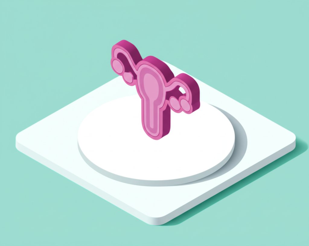
The Amazing Shrinking Uterus
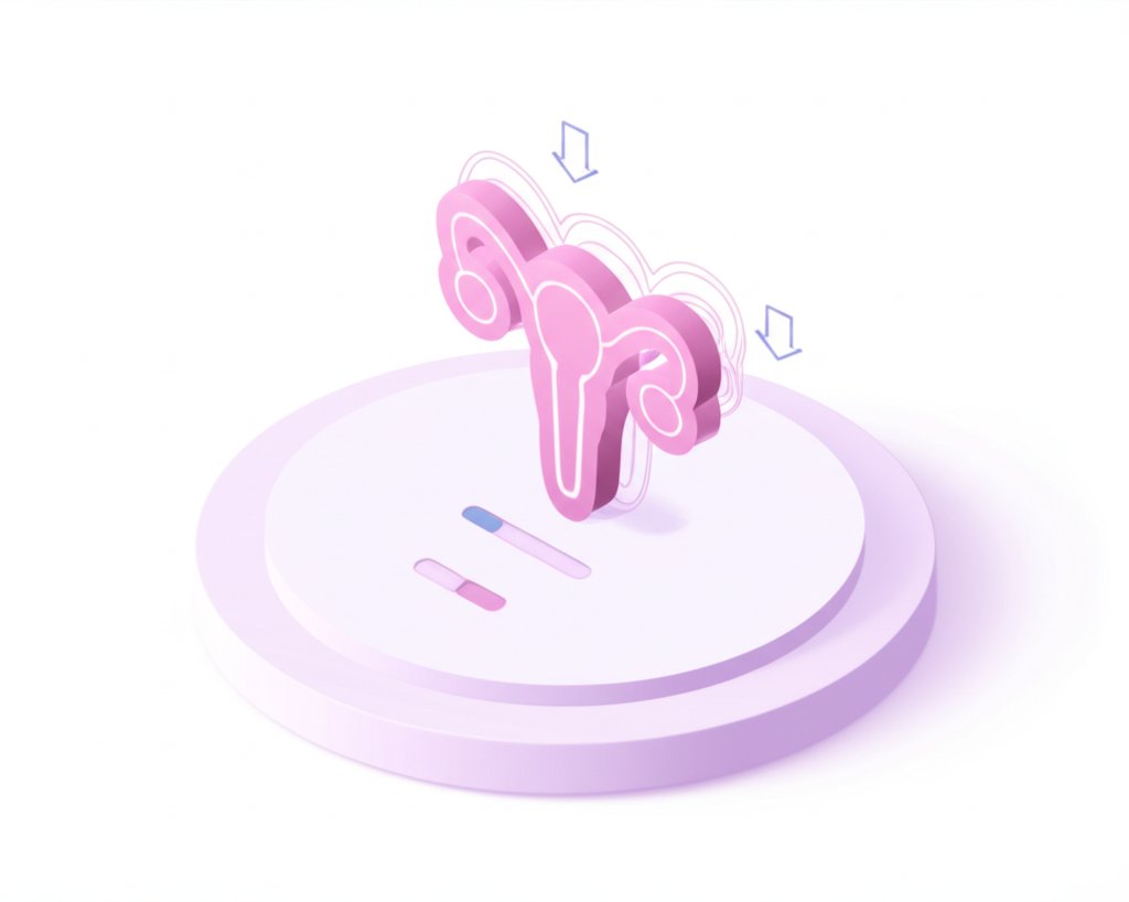
After delivery, your uterus embarks on a remarkable transformation, gradually returning to its pre-pregnancy size over approximately six weeks. This process, called involution, is unique to each woman, with some experiencing a quicker recovery than others. Immediately after birth, the uterus begins contracting to expel the placenta and control bleeding. These contractions, along with the shedding of the uterine lining (lochia), are essential aspects of **uterus involution**. The hormone oxytocin, released during breastfeeding, further stimulates these contractions, sometimes causing cramping known as “afterpains.” While potentially uncomfortable, afterpains signify that the uterus is actively healing. Over time, your belly will shrink and your uterus will feel firmer.
The First 48 Hours: A Critical Period
The first 48 hours postpartum are particularly crucial for uterine involution.1 During this time, the uterus undergoes rapid changes in size and position, moving higher in the abdomen, making examination slightly more challenging. This rapid shift underscores the importance of close monitoring during this initial period. Similar to muscle soreness after exercise, the uterus experiences its own version of “post-workout” discomfort—afterpains. Accompanying these cramps is lochia, a normal discharge composed of blood, mucus, and tissue from the healing uterine lining. The amount and color of lochia gradually decrease over the following weeks. Several factors influence the pace of involution, including the number of previous pregnancies (parity), delivery method (vaginal or cesarean), breastfeeding, and the baby’s size. Breastfeeding, for instance, often facilitates more efficient contractions due to the release of oxytocin. Healthcare providers carefully monitor uterine involution during postpartum checkups, using palpation and sometimes ultrasound.
Clinical Monitoring: Assessing the Involution of Your Uterus
Monitoring the rate of **involution of uterus** is a key part of postpartum care. Healthcare providers often clinically assess this by noting the height of the fundus (the top part of the uterus) in relation to the symphysis pubis (the joint at the front of your pelvis). This measurement should be taken carefully at a fixed time each day, ideally by the same observer, to ensure consistency. To secure an accurate reading, it’s crucial that the bladder is emptied beforehand, and preferably the bowel too, as a full bladder or loaded bowel can falsely elevate the fundal height. The uterus is gently centralized, and a measuring tape is used to determine the fundal height above the symphysis pubis. Immediately after delivery, the fundus typically lies just below or at the level of the umbilicus, gradually descending back into the pelvis over the subsequent weeks as **uterus involution** progresses.
Understanding the Anatomical and Physiological Journey of Uterine Involution
The process of **uterine involution** involves profound anatomical and physiological transformations. Beyond simply shrinking, the uterus undergoes a complex restructuring to restore its non-pregnant state.
Anatomical Transformations
- Uterus Size and Weight: Immediately after delivery, the uterus is a firm, retracted organ, measuring approximately 20x12x7.5 cm (length, breadth, thickness) and weighing about 1000 grams. By the end of six weeks, its dimensions are nearly identical to the non-pregnant state, and its weight significantly reduces to around 60 grams.
- Lower Uterine Segment: Post-delivery, the lower uterine segment, which became thin and stretched during pregnancy, transforms into a flabby, collapsed structure. It gradually reverts to its normal shape and size, becoming the part of the uterus between the body and the internal opening of the cervix over a few weeks.
- Cervix: Following childbirth, the cervix remains soft and open, admitting several fingers. However, as the `involution of uterus` progresses, it rapidly narrows. By the end of the first week, it typically admits only the tip of a finger as it closes back to its pre-pregnant form.
Physiological Processes Within the Uterus
- Muscles: During pregnancy, the muscle fibers of the uterus undergo marked hypertrophy (enlargement) and hyperplasia (increase in number), with individual fibers expanding up to 10 times in length and 5 times in breadth. During puerperium, the number of muscle fibers does not significantly decrease, but there is a substantial reduction in the size of these myometrial cells, contributing to the overall shrinkage of the uterus.
- Blood Vessels: Significant changes occur in the blood vessels, particularly at the placental site. The arteries constrict due to the contraction of the uterus and thickening of the intima, followed by thrombosis (blood clot formation). New, smaller blood vessels gradually grow inside these thrombi as the uterus heals.
- Endometrium: Following delivery, the majority of the decidua (the uterine lining during pregnancy) is shed with the expulsion of the placenta and membranes, especially at the placental site. The remaining endometrium, ranging from 2-5 mm in thickness, regenerates from the epithelium of the uterine gland mouths and interglandular stromal cells. Regeneration of the general uterine epithelium is typically completed by the 10th day postpartum, and the entire endometrium is restored by day 16, though the placental site takes about six weeks to fully heal.
Potential Complications and Ongoing Research
Although **uterus involution** usually progresses smoothly, complications like postpartum hemorrhage (heavy bleeding) or infection can occasionally occur. Retained placental fragments or subinvolution (delayed or incomplete involution) may also arise.2 Regular postpartum checkups are crucial for identifying and addressing any concerns. Contact your healthcare provider immediately if you experience heavy bleeding (soaking a pad every hour), persistent pain, or foul-smelling discharge. Ongoing research continues to explore the intricacies of involution, such as the specific mechanisms governing the process and the influence of factors like genetics and pre-existing health conditions. This ongoing research suggests our understanding of the process may continue to evolve, further enhancing our knowledge of the **involution of uterus**.
Involution vs. Puerperium: Understanding the Difference
While involution specifically refers to the uterus returning to its pre-pregnancy state, puerperium encompasses all the maternal physiological changes during the postpartum period. This broader term, often called the “fourth trimester,” typically lasts six to eight weeks, although some changes may persist longer. The puerperium is arbitrarily divided into three stages: **Immediate Puerperium** (within 48 hours post-delivery), **Early Puerperium** (up to 7 days), and **Remote Puerperium** (up to 6 weeks). The puerperium involves significant hormonal shifts, physical healing, and emotional adjustments. Key hormones like estrogen and progesterone decrease, while prolactin, responsible for milk production, increases. This hormonal fluctuation can cause various symptoms, from night sweats to mood swings. In addition to involution, the puerperium includes changes like decreased vaginal bleeding, perineal healing (after vaginal birth), and fluctuating energy levels. Emotional adjustments to life with a newborn are also a significant aspect of this period. While puerperium is a natural process, potential complications like postpartum hemorrhage, infections, pre-eclampsia, and postpartum depression can occur. Regular checkups are vital for early detection and management.
How Long Does Uterine Shrinkage Take?

Uterine involution, the process of the uterus shrinking back to its normal size, typically takes six to eight weeks. This process, which begins immediately after childbirth, can be likened to a balloon deflating, albeit a more complex and impressive one. Uterine muscles contract, returning to their pre-pregnancy shape, while the body sheds excess tissue and lining, contributing to postpartum bleeding (lochia). You’ll likely notice a gradual decrease in belly size, a firmer uterus, and changing lochia (from bright red to brownish, then yellowish-white). Cramping, known as afterpains, is also common, especially with subsequent pregnancies. Factors like breastfeeding can influence the speed of **uterus involution**, as the hormone oxytocin, released during nursing, stimulates uterine contractions. Multiple births may also slightly prolong the involution period.
Embarking on Your Postpartum Recovery
While six to eight weeks is the general timeframe for the **involution of uterus**, each woman’s experience is unique. Certain signs warrant immediate medical attention, such as heavy bleeding (soaking more than a couple of pads per hour), high fever (100.4°F/38°C or higher), or persistent pain unresponsive to over-the-counter medication. Ongoing research continues to investigate the specifics of uterine involution and its influencing factors. Remember, your body is undergoing a remarkable transformation. Practice patience, listen to your body, and reach out to your healthcare provider with any concerns. They are your best resource for support and guidance during this incredible postpartum journey. For further exploration into the body’s intricate anatomy, consider learning about the intertrochanteric crest, a crucial landmark that anchors essential muscular structures and shapes human locomotion.
Key Points to Remember:
- Involution of Uterus: The uterus shrinking back to its pre-pregnancy size and state after childbirth.
- Duration: Usually 6-8 weeks, but varies.
- Mechanism: Uterine contractions, hormonal changes, and tissue breakdown.
- Signs: Decreasing belly size, firmer uterus, vaginal discharge (lochia), afterpains.
- Potential Issues: Subinvolution, postpartum hemorrhage, infection.
- Importance of Checkups: Monitor involution and address concerns, including fundal height assessment.
- Warning Signs: Heavy bleeding, fever, persistent pain, foul-smelling discharge.
- Recovery: Patience, nutrition, hydration, and rest are crucial.
- Ongoing Research: The scientific understanding of postpartum recovery and **uterus involution** is constantly evolving.
1ScienceDirect Excerpt: “If the cow is less than 48 hours post partum, a hand may enter the uterus easily, but rapid cervical involution makes this difficult after 48 hours.” (This analogy helps illustrate the rapid changes occurring in the cervix during this period, although human anatomy differs.)
2 Delayed or incomplete involution can increase the risk of complications such as postpartum hemorrhage or infection.


1 thought on “Understanding Uterine Involution: Your Postpartum Journey”
Comments are closed.